The power to see more than the human eye can perceive, on the other hand, has far more practical applications than the heroic fantasies of a comic book fan. To figure out what’s wrong, doctors and healthcare experts frequently need to look inside the human body. Diagnostic imaging examinations, fortunately, can perform what the eye cannot. Radiologic technologists use their “powers” to help save lives—all without wearing a cape!
Diagnostic imaging tests that are commonly used
The most common diagnostic imaging exam performed in medical institutions is the X-ray, which is a wide term that covers a range of sub-categories. X-rays are used for a variety of purposes, including diagnosing the cause of pain, determining the severity of an injury, monitoring disease progression, and assessing the effectiveness of medicines.
CT scans
CT scans, also known as computed axial tomography scans or CAT scans, are a type of imaging that allows clinicians to observe cross-sections of the body. In comparison to a traditional X-ray, cross-sectional pictures produce more detailed images. In fact, when something suspect appears on an X-ray, a CT scan is frequently requested.
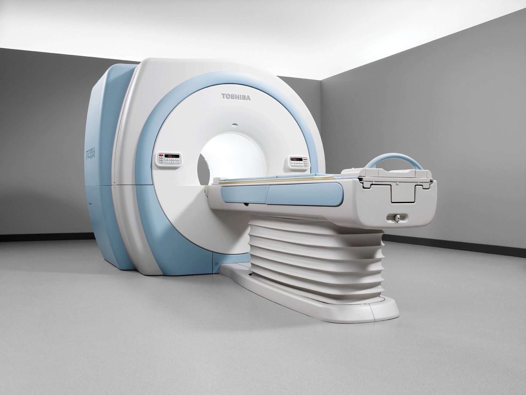
MRI
Magnetic resonance imaging, or MRI, is another option for cross-sectional imaging. Soft tissues, such as organs and tendons, are imaged with MRIs, which are similar to CAT scans. neuro MRI in New Jersey, unlike CAT scans, employ radio waves and magnetic fields rather than ionizing radiation. Because MRIs do not involve radiation, they are often seen to be safer, but they also take longer to perform.
Mammogram
In the fight against breast cancer, early diagnosis is critical. There are two forms of mammography: screening and diagnostic mammograms. Mammography screening is done to see if there are any abnormalities in the first place. Diagnostic mammography is performed to examine for cancer once a tumor or thickening in the breast is identified.
Ultrasound
Ultrasound, also known as sonography, uses high-frequency sound waves to acquire images from within the body. It’s frequently used to look for problems with soft tissues like organs and arteries. Ultrasounds are the preferred method of examining pregnant women because they do not use radiation.
Fluoroscopy
Fluoroscopy is like a motion picture of bodily functions, whereas other examinations are like still photography. Contrast dyes are frequently used in the operation to illustrate how they travel through the body. An X-ray beam provides signals to a monitor while all of this is going on. Fluoroscopies are used to examine both hard and soft tissue, such as bones, joints, organs, and blood vessels.
PET scans
A PET scan, also known as a positron emission tomography scan, is a type of body scan that detects disease at the cellular level. Radioactive tracers are injected into the body during the process. The tracers employ a PET scanner to spot issues that could otherwise go undiscovered until they worsen.